Prairie Heart at St. Mary's combines advanced technology and innovative treatment with a caring tradition, providing quality health care close to home. With access to more than 117 heart specialists, the doctors at Prairie Heart Institute provide the highest quality of heart care from diagnostic services to treatment to rehabilitation.
Heart and Vascular Care
Prairie Heart Institute at HSHS St. Mary's Hospital delivers state-of-the-art services with the most compassionate care — from prevention and diagnosis to treatment and recovery.
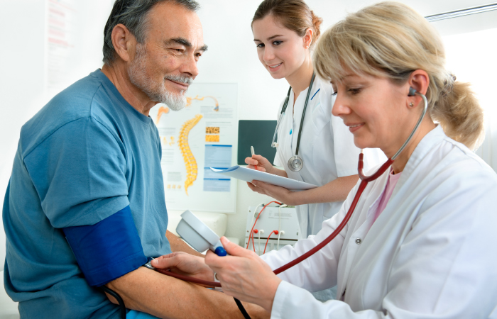
Screenings and tests
Identifying a person's risk of heart disease with early detection
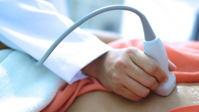
Echocardiogram (ECHO)
Ultrasound is one of the most widely used diagnostic procedures available. It provides a safe, non-invasive and virtually painless means of observing soft tissue anatomy on an outpatient basis.
An echocardiogram is a safe, non-invasive ultrasound imaging procedure used to assess cardiac function. Echocardiography allows doctors to visualize the anatomy, structure, and function of the heart. The echocardiogram can show:
An echocardiogram is a safe, non-invasive ultrasound imaging procedure used to assess cardiac function. Echocardiography allows doctors to visualize the anatomy, structure, and function of the heart. The echocardiogram can show:
- all four chambers of the heart
- the heart valves
- the blood vessels entering leaving the heart
- the sack around the heart.
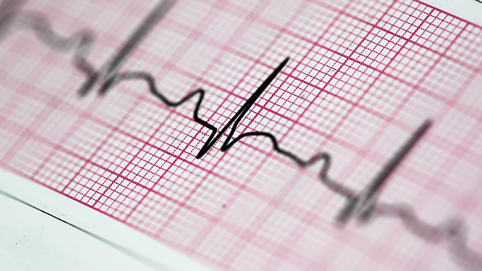
Electrocardiogram (EKG)
The electrocardiogram is a device capable of recording the electrical activity of the heart from electrodes placed on the skin in specific locations.
Physicians use this information to discover such things as:
Physicians use this information to discover such things as:
- heart rate.
- arrhythmias.
- myocardial infarctions.
- atrial enlargements.
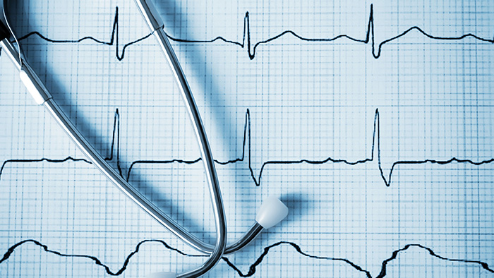
Electrophysiology for Arrythmias
Electrophysiology is a specialized branch of cardiovascular care that concentrates on heart arrhythmias. Heart arrhythmias include a number of cardiac conditions that affect the heart’s ability to maintain a regular beat. Some may cause the heart to beat too slowly or too fast, both with potentially serious implications. Left undiagnosed, arrhythmias can contribute to a heart attack.
Electrophysiologists with the Prairie Heart Institute, provide specialized electrophysiology procedures at HSHS St. Mary’s Hospital. During a diagnostic electrophysiology test, a thin electrode is inserted into a vein in your arm or groin area. Once inserted, the electrode is moved to your heart and used to trigger controlled arrhythmias. The electrical signals of the heart are then recorded, identified so that the location of the disorder can be determined. Knowing how your heart reacts during an arrhythmia allows the physician to provide more focused care.
Many arrhythmias are treated through the insertion of a pacemaker. A pacemaker is a small device, weighing about an ounce. It uses electrical impulses to help regulate the beating of the heart.
Heart monitoring
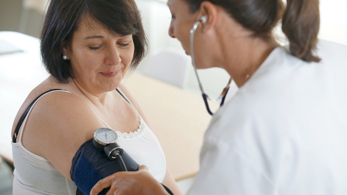
Blood Pressure Monitoring
A blood pressure monitor consists of a blood pressure cuff connected to a monitor. Blood pressures are taken hourly for a 24 hour period. Physicians then utilize the information to determine hypertension (high blood pressure). This requires a 30 minute appointment.
Holter Monitor
A holter monitor is worn for a 24 or 48 hour period during which the electrical activity of the heart is recorded. The recording is read and the information reported is used by your physician to determine what medical treatment is needed, if any. The monitor is connected to the chese with 5 patches and wires. This requires a 30 minute appointment.
Event Monitor
An event monitor is issued for a 30 day period. Two patches are applied to the chest and connected by wires to a small transmitter. Patches are changed daily by the patient, allowing for bathing. Over a 30 day period, the patient takes a recording when an event or episode occurs. The recording is then transferred via a landline phone to the monitor lab. Physicians utilize this information for heart rhythm abnormalities. This requires a 30 minute appointment.
Stress tests
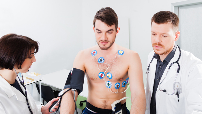
A stress test, sometimes called a treadmill test or nuclear stress test, helps determine how well the heart handles work. As the patient walks on a treadmill or is given medication to stress the heart, an EKG machine monitors the heart rate, breathing, blood pressure, the electrical activity of the heart, and exercise tolerance. With a nuclear stress test, there are two parts to the test: a resting study and a stress study
A Nuclear Stress Test is used by doctors to diagnose and monitor heart disease.
During a Nuclear Stress Test, a safe amount of a radioactive drug is injected into your vein which allows the cardiologist to see how well blood is flowing to and through your heart.
The test, usually conducted over two days, consists of taking images of your heart in two phases: a stress phase and a resting phase.
There may be restrictions as you prepare for this test. Please ask your health care provider.
