- X-ray
- Radiographer/Fluoroscopy
- MRI (in-house)
- Nuclear Medicine
- 64-slice CT Scanner (completes a full body scan with seconds!)
- Ultrasound
- 3-D Mammography
Imaging
Advanced imaging technology at HSHS Holy Family Hospital
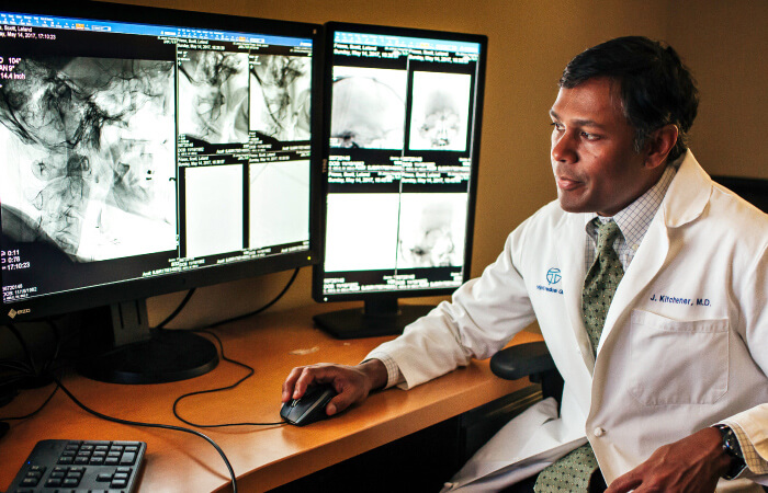
Learn more
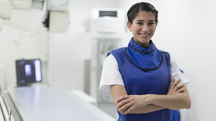
X-Rays
X-ray, commonly referred to as radiography, uses ionizing radiation to provide images of the body. Radiography is used in many ways to diagnose disease and injuries. Some common exams are X-rays, GI fluoroscopy exams and joint injections (arthrograms). Preparation for this scan varies by patient based on the test being conducted.

3D mammography
HSHS Holy Family Hospital is transforming breast cancer screening and detection with the latest 3D mammography technology and a focus on your comfort. Our goal is to make breast health as easy as 1, 2, 3.
Instead of reviewing the breast tissue in a single, flat image, this technology folds together a series of images to create a 3D view, allowing doctors to see the tiniest details and find cancer in its earliest stages.
Learn more.
Instead of reviewing the breast tissue in a single, flat image, this technology folds together a series of images to create a 3D view, allowing doctors to see the tiniest details and find cancer in its earliest stages.
Learn more.
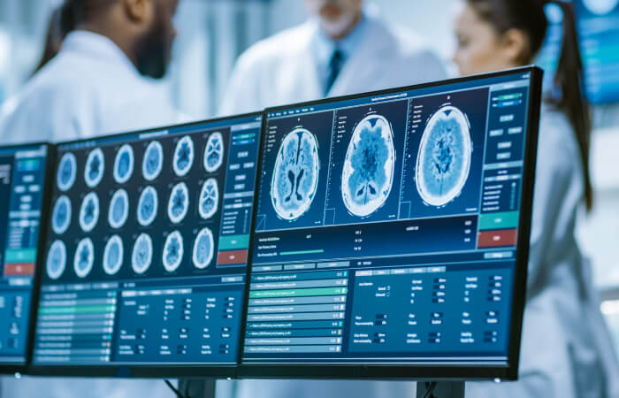
CT scan (computerized tomography)
During this test, the patient lies in a doughnut-shaped machine that takes pictures of the body. The scanner is used in combination with a digital computer to create "slices" of different organs of the body, making it possible to detect diseases sooner than with a regular X-ray. CT scans are conducted for calcium scoring at the Prevea Allouez Health Center.
To prepare for a CT scan
Wear loose, comfortable clothing free of metal snaps and zippers, if possible. When you arrive, you will be asked to remove metallic jewelry, watches, hair pins, hearing aids, removable dental work and glasses if they are in the area being scanned.
If you are having a CT scan of the abdominal area, you may need to drink a liquid contrast. This will be given to you, with instructions, in advance. You may be asked to not eat or drink anything for four hours before your scan. Your health care provider will instruct you which medications, if any, cannot be taken.
The scan typically takes 30 minutes or less. You will be asked to hold very still in specific positions as instructed by a technologist. You may be asked to hold your breath during some scans to reduce blurring on the images. If your scan requires the use of a contrast, you may be given another contrast material during the scan by IV injection. This may make you feel warm inside, but the sensation only lasts a few moments and is not painful. After the scan, you will be asked to drink plenty of fluids.
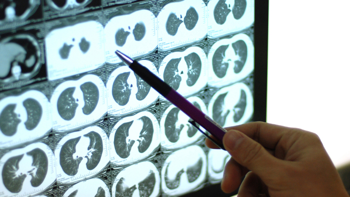
Lung cancer screening
Are you one of the thousands of Americans who is at risk for developing lung cancer? According to the American Cancer Society, more people die of lung cancer each year than of colon, breast, and prostate cancers combined. The good news is that when diagnosed early, lives can be saved. HSHS Holy Family Hospital's Lung Cancer Screening can do just that.
Holy Family's Lung Cancer Screening is a quick, painless, non-invasive low dose CT scan which can detect nodules or spots on your lung which might be early indicators of lung cancer. It takes just one-minute to have a scan which can potentially save your life.
Lung cancer screenings are most beneficial to those with the highest risk of lung cancer:
A physician’s order is required for a Lung Cancer Screening. If you would like to consider having this screening, please make an appointment with your family physician to have an informed discussion on the potential benefits, limitations and possible risks of having a Lung Cancer Screening. After reviewing and discussing the criteria for the Lung Cancer Screening, your physician will determine if you are a candidate for a Lung Cancer Screening.
Holy Family's Lung Cancer Screening is a quick, painless, non-invasive low dose CT scan which can detect nodules or spots on your lung which might be early indicators of lung cancer. It takes just one-minute to have a scan which can potentially save your life.
Lung cancer screenings are most beneficial to those with the highest risk of lung cancer:
- Current or former smokers who are older: A lung cancer screening is generally an option for smokers and former smokers who are 50 years or older. Check with your insurance provider or Medicare on their age/smoking history requirement, as well as any other criteria.
- Smoked heavily for many years: Those who have a tobacco smoking history of at least 20 “pack years” – an average of one pack (20 cigarettes) per day for 20 years.
- Once smoked heavily but have since quit: Those who were once a heavy smoker for a long time but have since quit smoking in the last 15 years.
A physician’s order is required for a Lung Cancer Screening. If you would like to consider having this screening, please make an appointment with your family physician to have an informed discussion on the potential benefits, limitations and possible risks of having a Lung Cancer Screening. After reviewing and discussing the criteria for the Lung Cancer Screening, your physician will determine if you are a candidate for a Lung Cancer Screening.
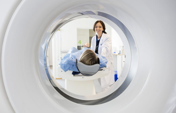
MRI (magnetic resonance imaging)
An MRI is a non-invasive and painless procedure in which radio waves and powerful magnets linked to a computer are used to create remarkably clear and detailed pictures of internal organs and tissues without the use of radiation.
To prepare for an MRI:
For your safety, you will be asked to put on a patient gown for the test. Clothing with metal or pockets will not be allowed in the scanning room. Certain types of metal in the area being scanned can cause significant errors, called artifacts, in the images. It is important that metal objects are not brought into the scanning room. You will be asked to remove your jewelry, watch, hairpins, bobby pins and hearing aids. A typical exam lasts between 30 to 60 minutes for each body area being scanned. You should allow extra time because the exam may last longer than expected.
The magnet makes a “knocking” sound as images are being taken. In between scans, the machine is quiet. The MRI technologist will provide you with either ear plugs or headphones for you to listen to music during your exam.
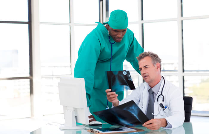
Nuclear Medicine
Nuclear medicine is a branch of medical imaging that uses small amounts of radioactive material to diagnose and determine the severity of, or treat, a variety of diseases, including cancers, heart disease, gastrointestinal, endocrine, skeletal and other abnormalities within the body.
Depending on the type of nuclear medicine exam, the radiotracer is injected into the body, swallowed or inhaled, and accumulates in the organ or area of the body being examined. Radioactive emissions from the radiotracer are detected by a special camera that produces pictures and provides information. Scanning can begin immediately after injection, or be delayed for several hours or even days. Scan time also varies, with some scans as short as 30 minutes and others several hours. Preparation for nuclear medicine exams varies, so instructions should be reviewed and followed carefully for each exam.
Depending on the type of nuclear medicine exam, the radiotracer is injected into the body, swallowed or inhaled, and accumulates in the organ or area of the body being examined. Radioactive emissions from the radiotracer are detected by a special camera that produces pictures and provides information. Scanning can begin immediately after injection, or be delayed for several hours or even days. Scan time also varies, with some scans as short as 30 minutes and others several hours. Preparation for nuclear medicine exams varies, so instructions should be reviewed and followed carefully for each exam.
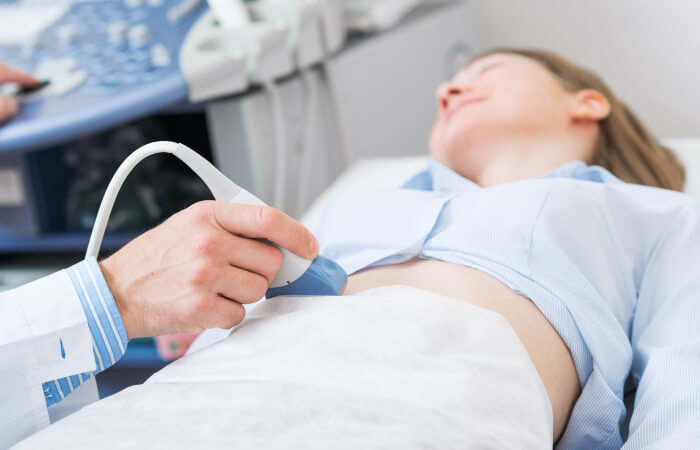
Ultrasound
During an ultrasound, you lie on an examination table and a clear gel is applied to the areas of the body being studied. This helps the transducer make firm contact with your body. It is then moved back and forth over the area of interest and uses high frequency sound waves to obtain images.
